Home

Géant salade comédie microscope 3d France utilisation Savant glucides

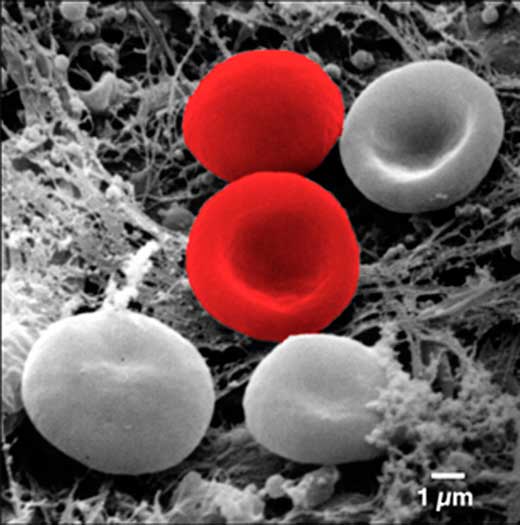
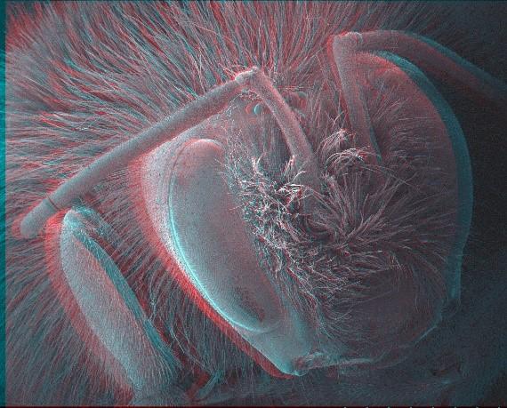
3D scanning electron microscopy for biology - Nanoanalysis - Oxford  Instruments
3D scanning electron microscopy for biology - Nanoanalysis - Oxford Instruments

Welcome
Welcome

Measuring Surface Roughness: The Benefits of Laser Confocal Microscopy |  Test & Measurement | Photonics Handbook | Photonics Marketplace
Measuring Surface Roughness: The Benefits of Laser Confocal Microscopy | Test & Measurement | Photonics Handbook | Photonics Marketplace
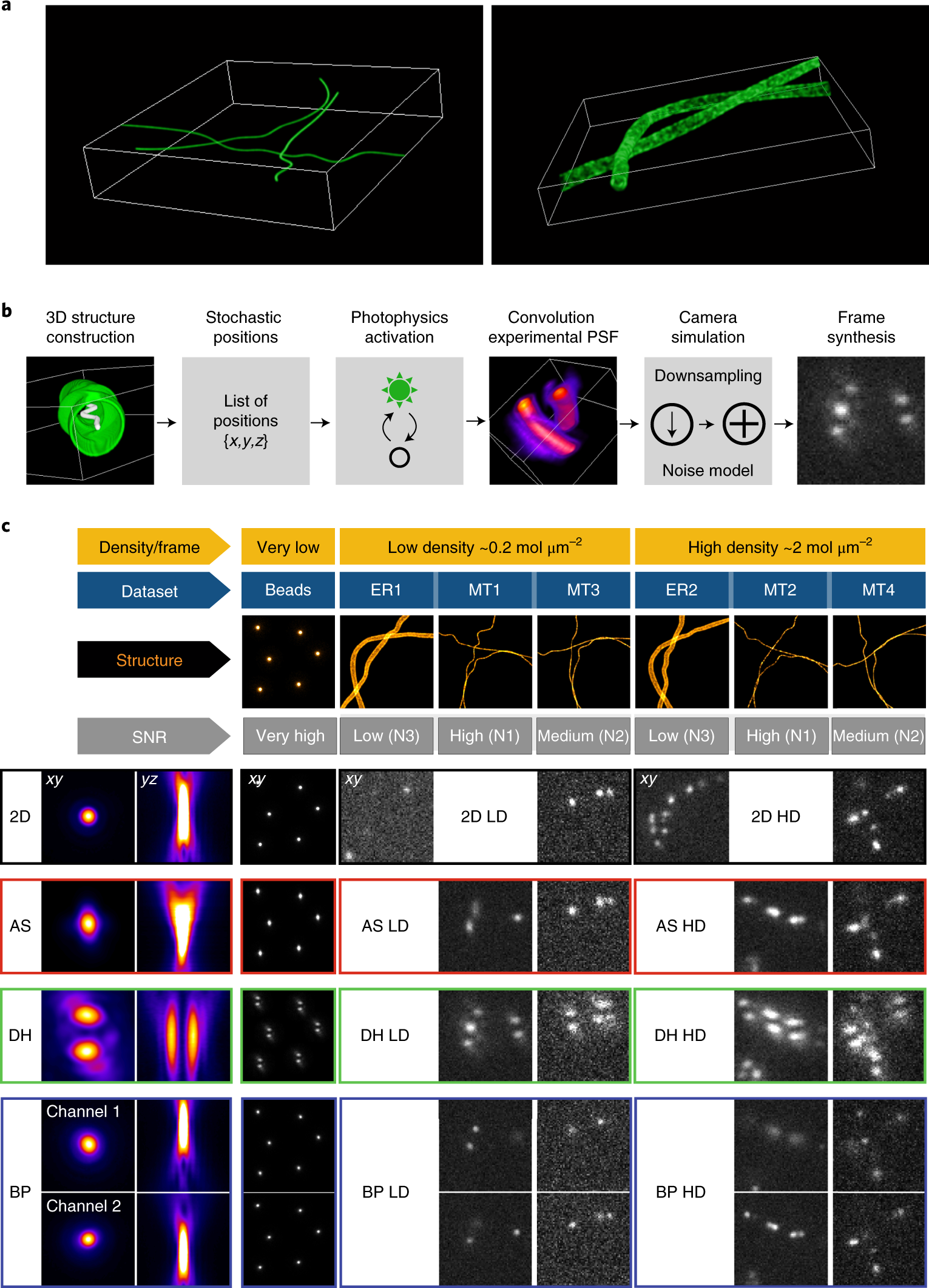
Super-resolution fight club: assessment of 2D and 3D single-molecule  localization microscopy software | Nature Methods
Super-resolution fight club: assessment of 2D and 3D single-molecule localization microscopy software | Nature Methods
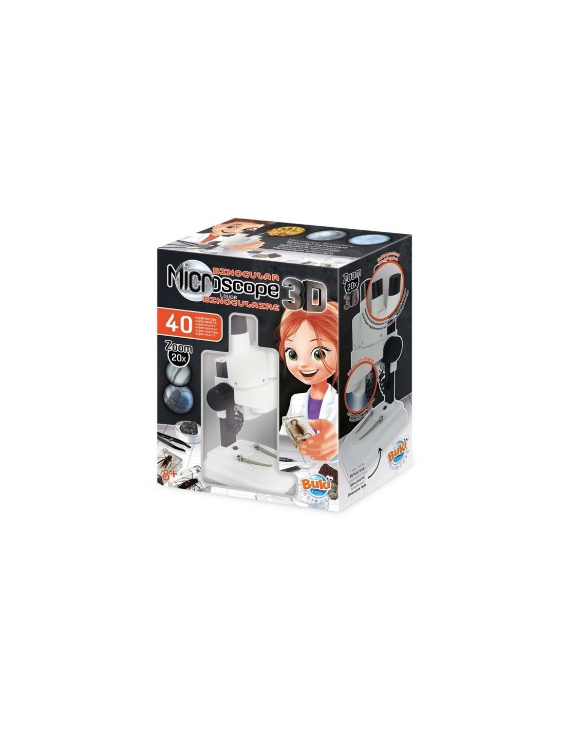
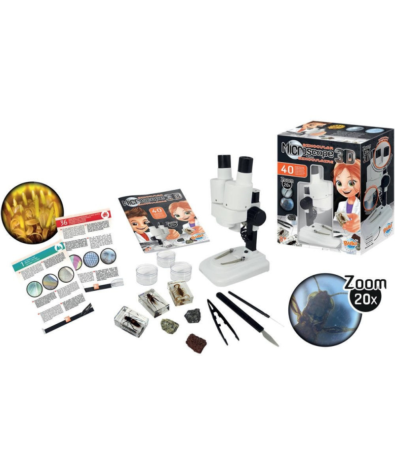
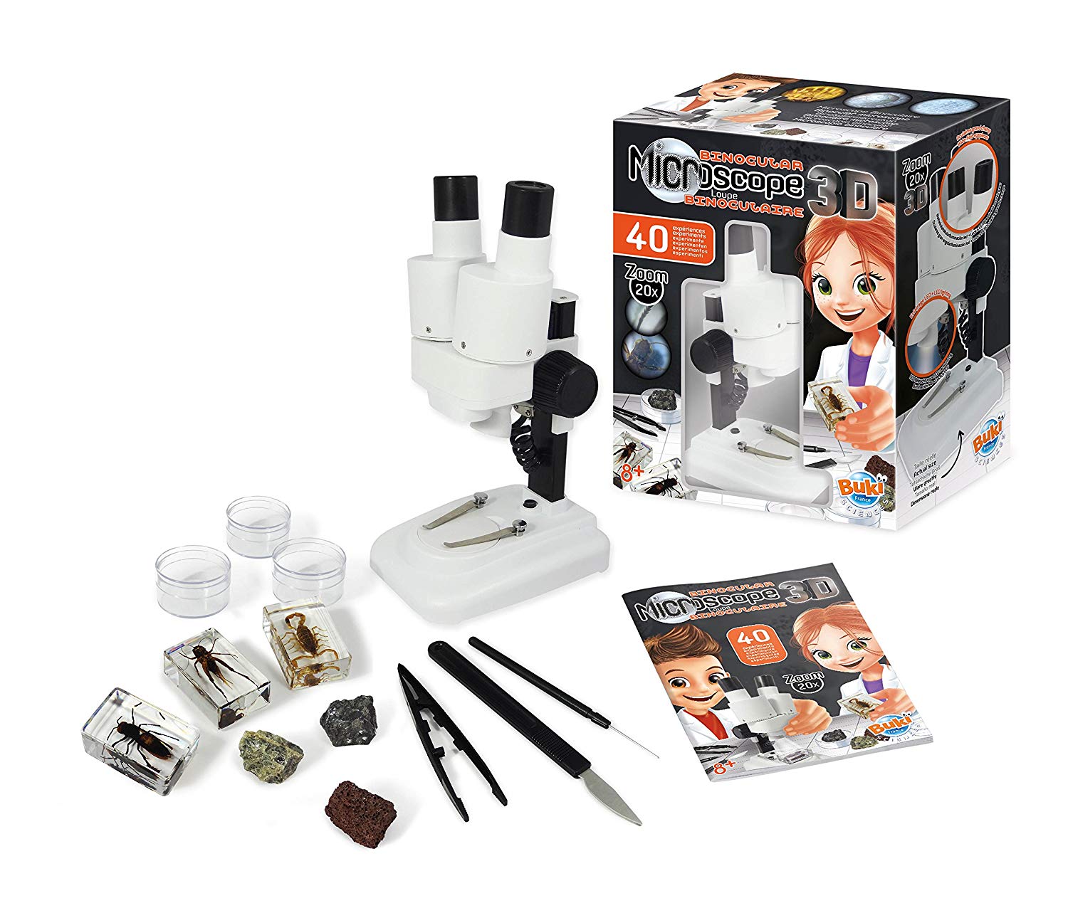
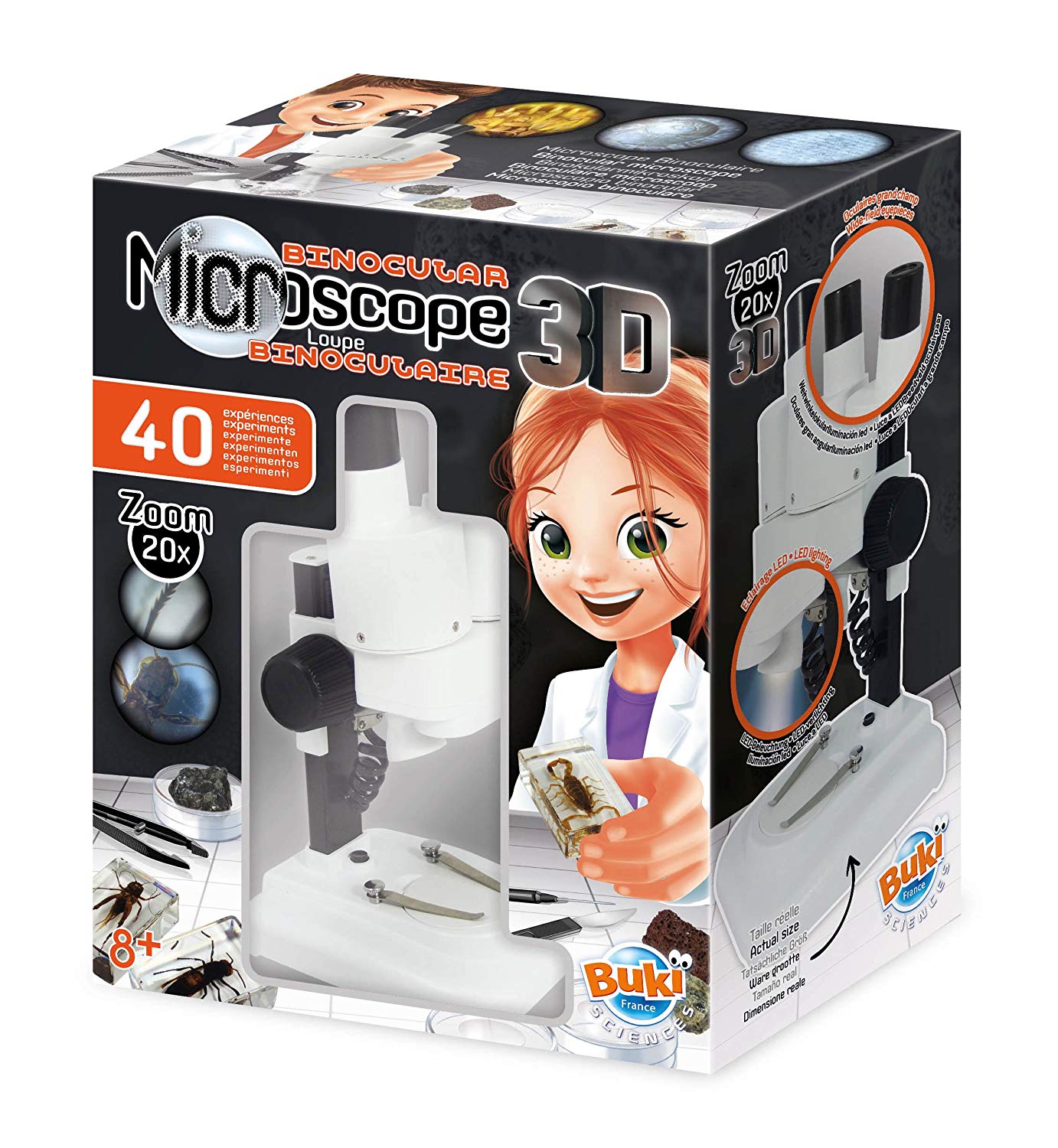
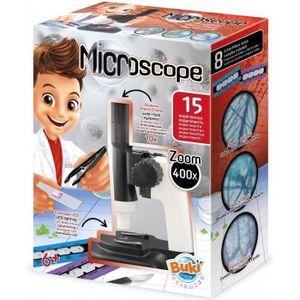
Microscope stereo 3D - Buki

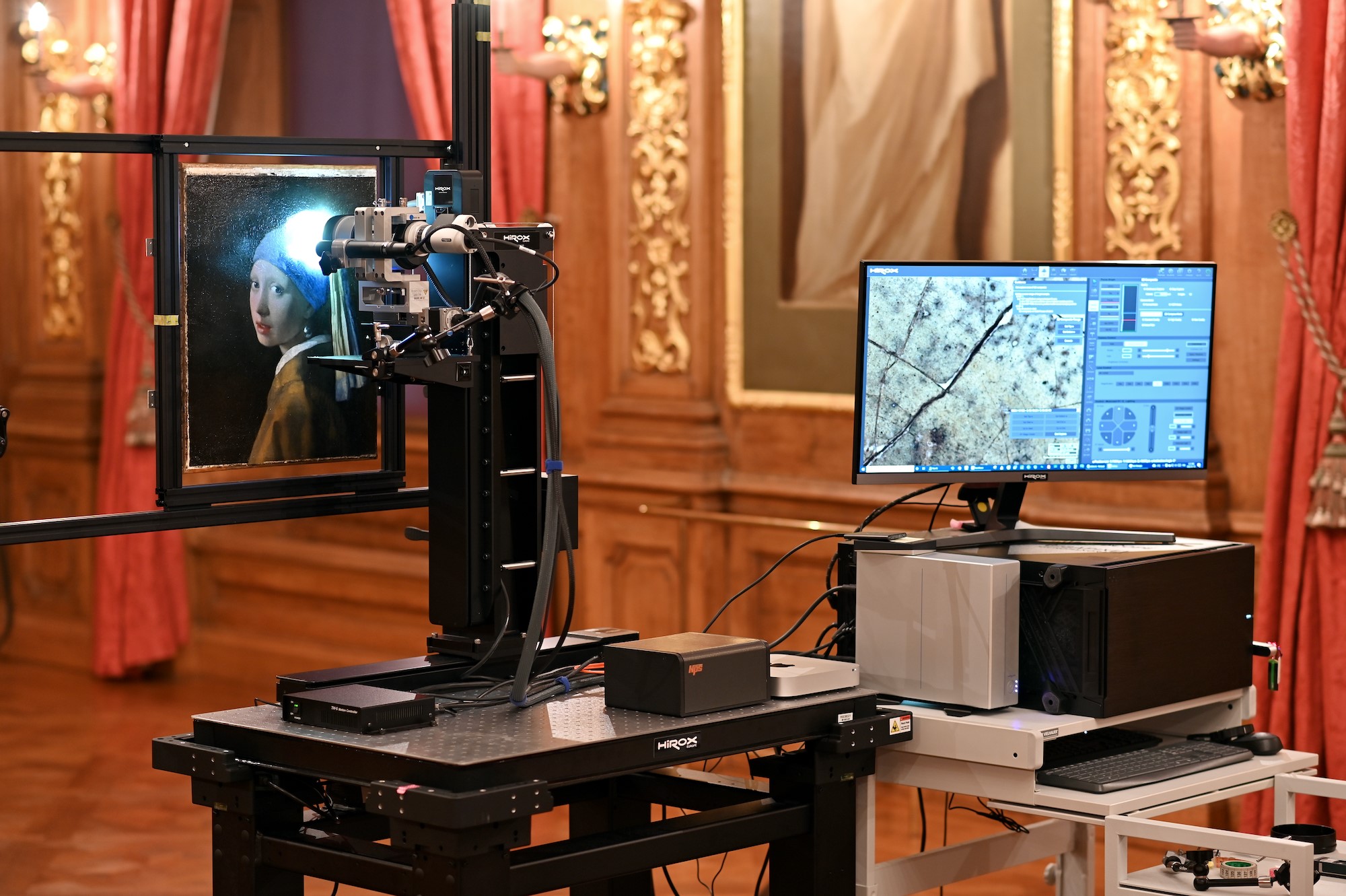
Hirox in Museums - HIROX 3D Digital Microscopy for Art and Conservation

PDF) Fast prototyping of 3D microniches with PRIMO

3D Res/volution event - FIB-SEM and Lattice SIM Elyra 7 - France-BioImaging

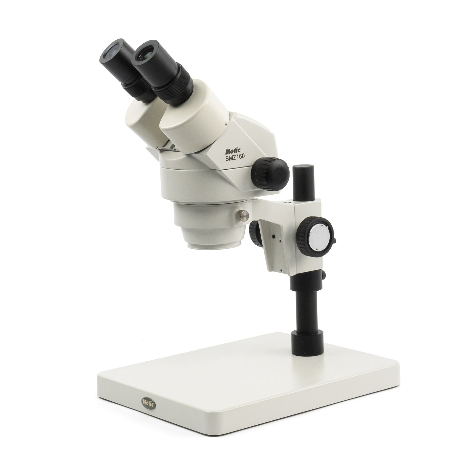
Motic Scientific
Motic Scientific

From 3D Light to 3D Electron Microscopy | Ebook

LensAFM — AFM for optical microscopes - Nanosurf

Microscopes – observe the living to fight disease - News from the Institut  Pasteur
Microscopes – observe the living to fight disease - News from the Institut Pasteur

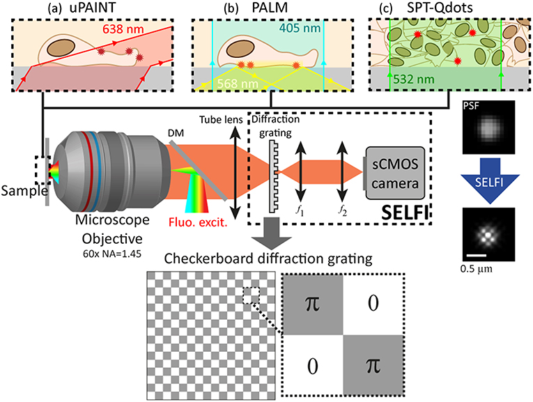
Frontiers | Self-Interference (SELFI) Microscopy for Live Super-Resolution  Imaging and Single Particle Tracking in 3D
Frontiers | Self-Interference (SELFI) Microscopy for Live Super-Resolution Imaging and Single Particle Tracking in 3D
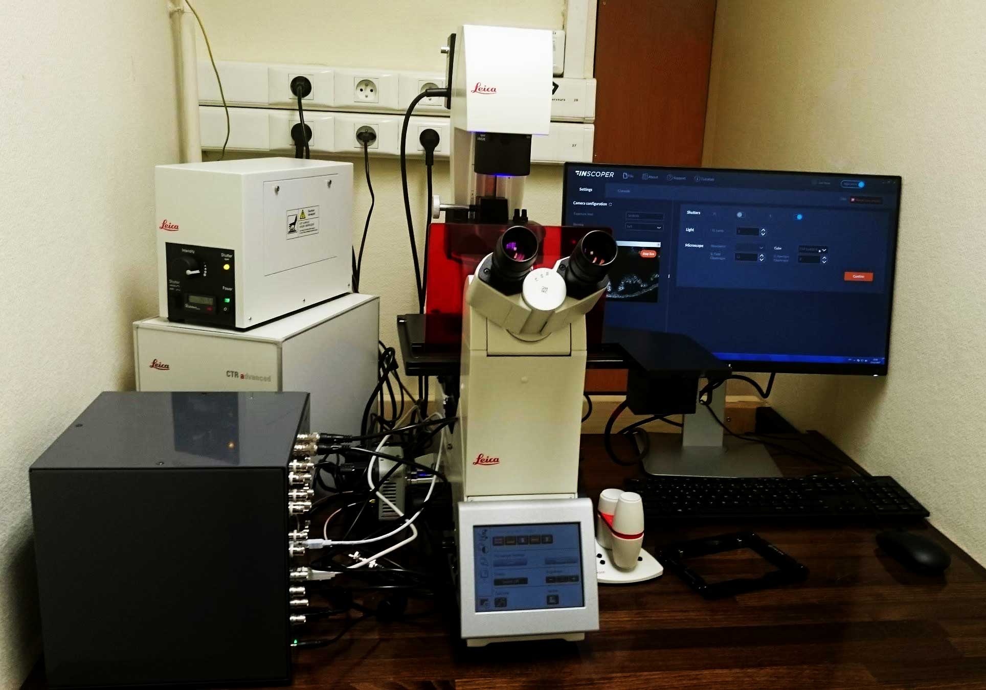
3D High Throughput Imaging - Nice, France - INSCOPER

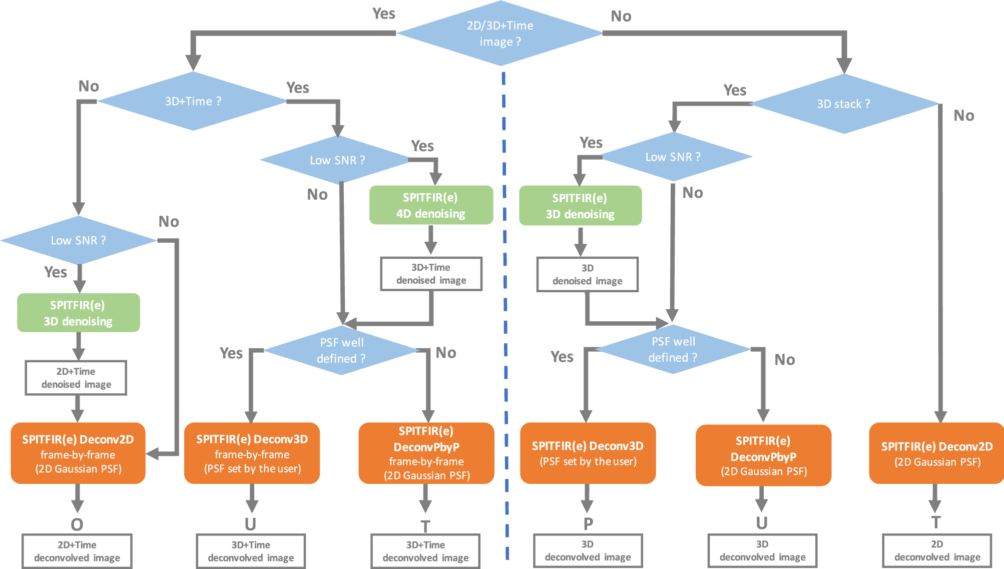
SPITFIR(e): a supermaneuverable algorithm for fast denoising and  deconvolution of 3D fluorescence microscopy images and videos | Scientific  Reports
SPITFIR(e): a supermaneuverable algorithm for fast denoising and deconvolution of 3D fluorescence microscopy images and videos | Scientific Reports

Compound Microscope and Case, Micrometric stage invented by  Michel-Ferdinand d'Albert d'Ailly, sixth duke of Chaulnes (French, 1714 -  1769), Gilt-bronze mounts attributed to Jacques Caffieri (French, 1678 -  1755 (master 1714)), Paris,
Compound Microscope and Case, Micrometric stage invented by Michel-Ferdinand d'Albert d'Ailly, sixth duke of Chaulnes (French, 1714 - 1769), Gilt-bronze mounts attributed to Jacques Caffieri (French, 1678 - 1755 (master 1714)), Paris,

3D Fluorescence Cell Imaging with Different Technologies.

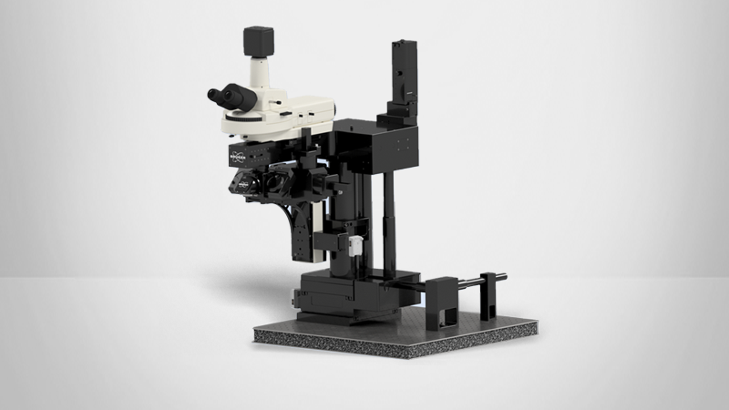
TruLive3D Imager | Bruker

SMZ-160 | Motic Microscopes

Transmission Electron Microscopy: Exploring the architecture of a virus in  all its forms - Workshop - France-BioImaging
Transmission Electron Microscopy: Exploring the architecture of a virus in all its forms - Workshop - France-BioImaging

From 3D Light to 3D Electron Microscopy | Ebook

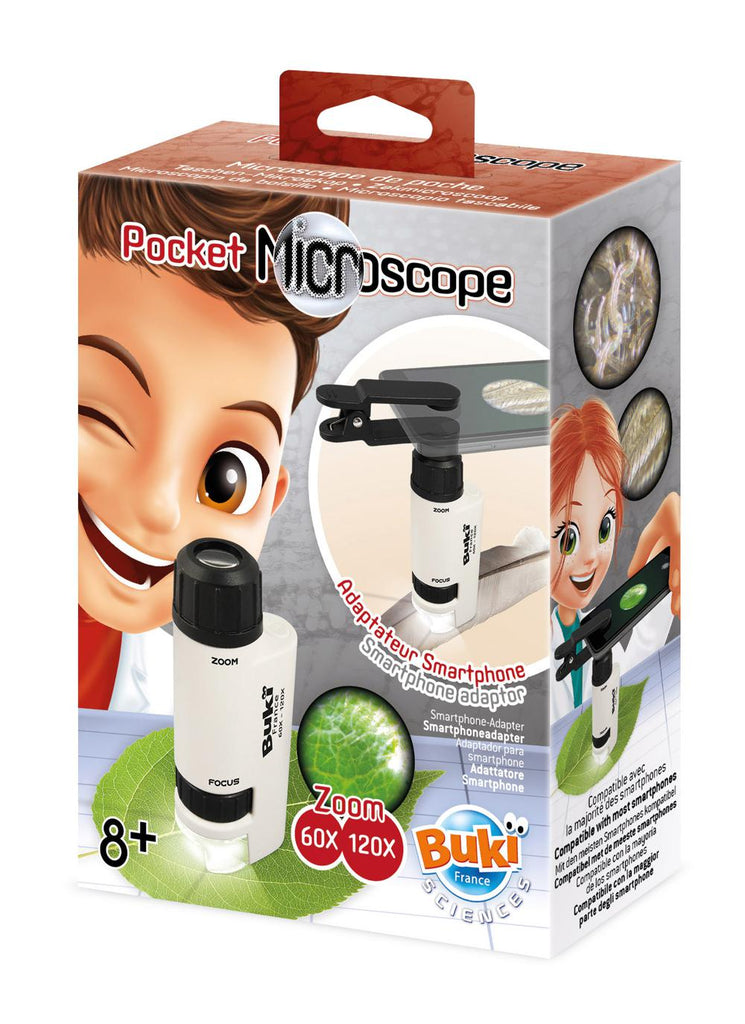
Buki France - Pocket microscope 120x – Touty Toys

MR700 Microscope 3 en 1 FR - Buki France - YouTube

BUKI - MICROSCOPE STEREO 3D MR500 - Achat - BUKI - MICROSCOPE STEREO 3D  MR500
BUKI - MICROSCOPE STEREO 3D MR500 - Achat - BUKI - MICROSCOPE STEREO 3D MR500

Buki France-Binocular microscope

What is Super-Resolution Microscopy? STED, SIM and STORM Explained |  Technology Networks
What is Super-Resolution Microscopy? STED, SIM and STORM Explained | Technology Networks

3D scanning electron microscopy for biology - Nanoanalysis - Oxford  Instruments
3D scanning electron microscopy for biology - Nanoanalysis - Oxford Instruments

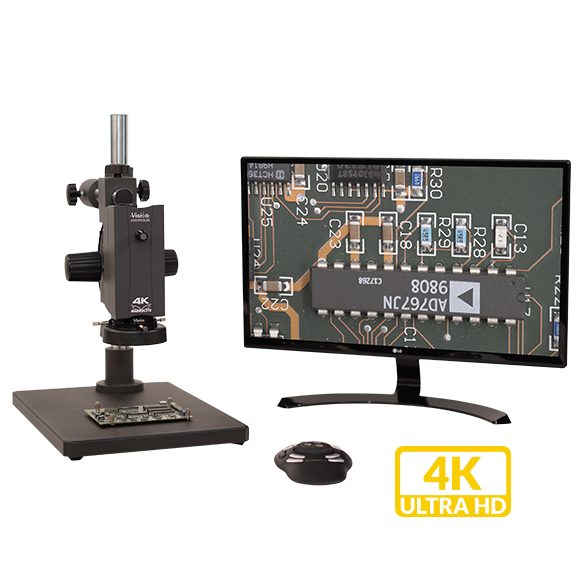
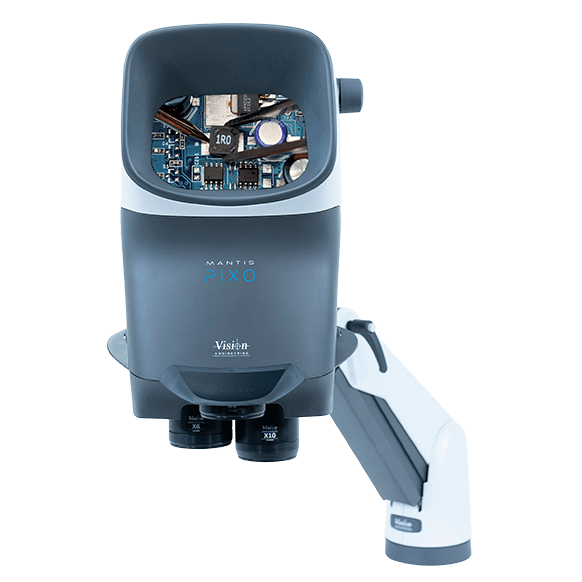
Makrolite 4K UHD digital microscope - Vision Engineering

3D Correlative Cryo-Structured Illumination Fluorescence and Soft X-ray  Microscopy Elucidates Reovirus Intracellular Release Pathway - ScienceDirect
3D Correlative Cryo-Structured Illumination Fluorescence and Soft X-ray Microscopy Elucidates Reovirus Intracellular Release Pathway - ScienceDirect

BUKI France MR500 – Binocular Microscope 3D – TopToy

3D Electron Microscopy : From Molecule to Organism : 22-23 May - France -BioImaging

Holotomography: 3D Non-Invasive Live-Cell Imaging of Stem Cells - PromoCell

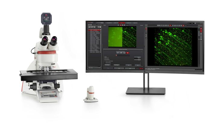
Microscope Cameras | Products | Leica Microsystems

Microscope Binoculaire - MR500 - BUKI France - YouTube

Color (and 3D) for Scanning Electron Microscopy | Microscopy Today |  Cambridge Core
Color (and 3D) for Scanning Electron Microscopy | Microscopy Today | Cambridge Core

KLS JCR 12V100WH10/5 OP-91641 12V100W KEYENCE 3D laser microscope light  bulbs | eBay
KLS JCR 12V100WH10/5 OP-91641 12V100W KEYENCE 3D laser microscope light bulbs | eBay

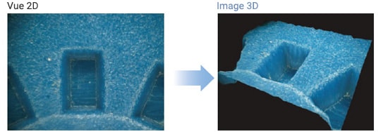
Capture d'image 3D | KEYENCE France

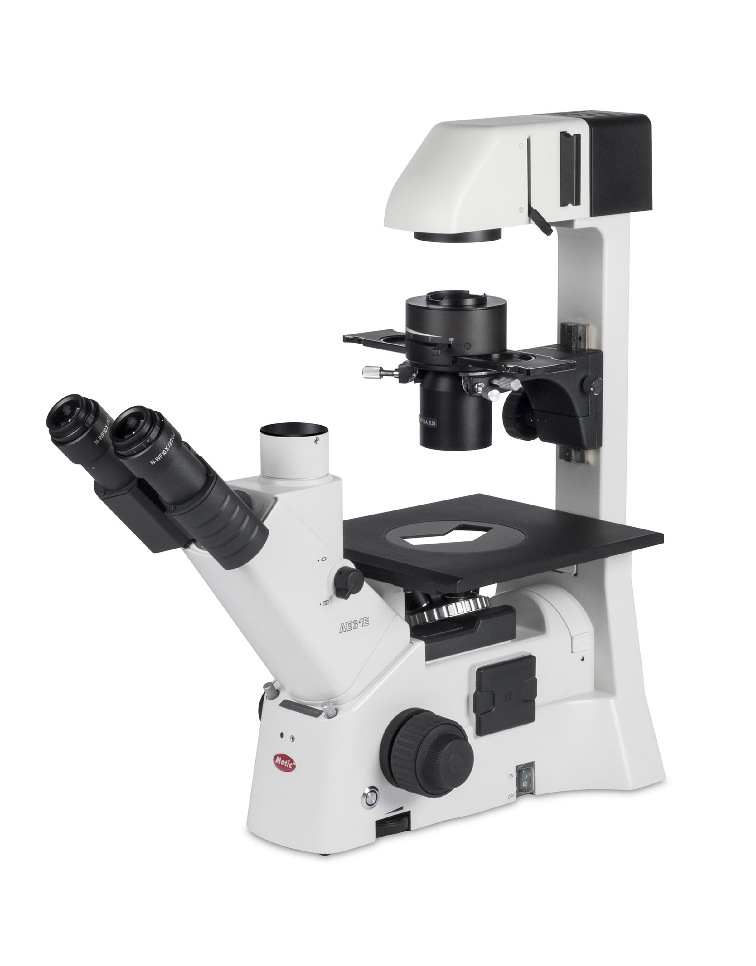
AE31 Elite | Motic Microscopes

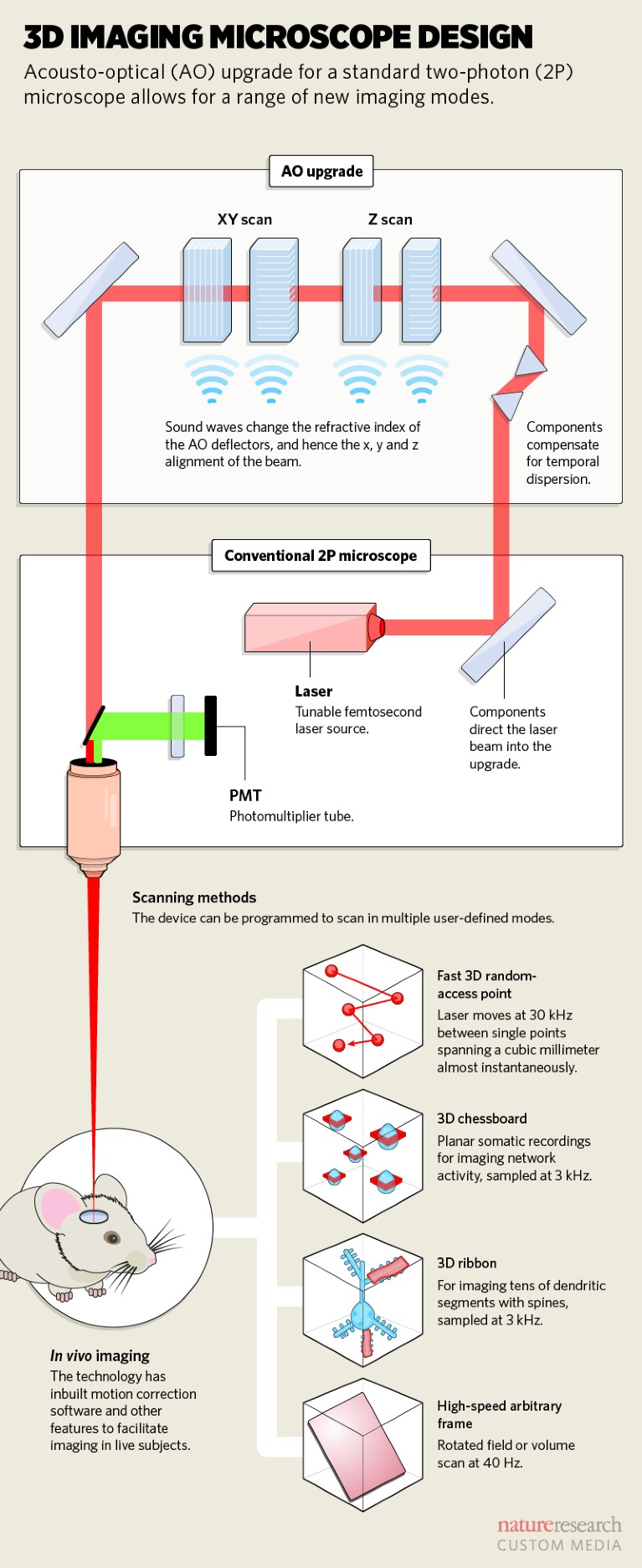
Sounding out the brain: acoustic signals focus light on neurons

3D scanning electron microscopy for biology - Nanoanalysis - Oxford  Instruments
3D scanning electron microscopy for biology - Nanoanalysis - Oxford Instruments

Zebrafish Research | Applications | Leica Microsystems

BUKI France MR500 – Binocular Microscope 3D – TopToy

Stereo Microscopes for 3D inspection - Vision Engineering

ZEISS Stemi 508 Stereo Microscope with 8:1 Zoom

Microscope on France Flag Background - Science Development Concept.  Research in Clinical Medicine or Biotechnology 3D Illustration Stock  Illustration - Illustration of nanotechnology, design: 203594734
Microscope on France Flag Background - Science Development Concept. Research in Clinical Medicine or Biotechnology 3D Illustration Stock Illustration - Illustration of nanotechnology, design: 203594734

Microscope stereo 3D - Buki - lapouleapois.fr - YouTube

Microscopy Technologies - France-BioImaging

Microscope Optico Paris /made in France /vintage - Etsy

Microscope 3d - Cdiscount

Microscope on France Flag Background - Science Development Concept.  Research in Clinical Medicine or Biotechnology 3D Illustration Stock  Illustration - Illustration of nanotechnology, design: 203594734
Microscope on France Flag Background - Science Development Concept. Research in Clinical Medicine or Biotechnology 3D Illustration Stock Illustration - Illustration of nanotechnology, design: 203594734

Un nouveau microscope confocal à la plateforme M&A ! - Réseau Mesure

Ultima Investigator Plus | Bruker

Habitium : Microscope 3D à lumière transmise et réfléchie avec rotation du  miroir à 360° sur son axe IVM 3D PCE Instruments
Habitium : Microscope 3D à lumière transmise et réfléchie avec rotation du miroir à 360° sur son axe IVM 3D PCE Instruments

Self-interference 3D super-resolution microscopy for deep tissue  investigations | Nature Methods
Self-interference 3D super-resolution microscopy for deep tissue investigations | Nature Methods

Stereo microscope - Wikipedia